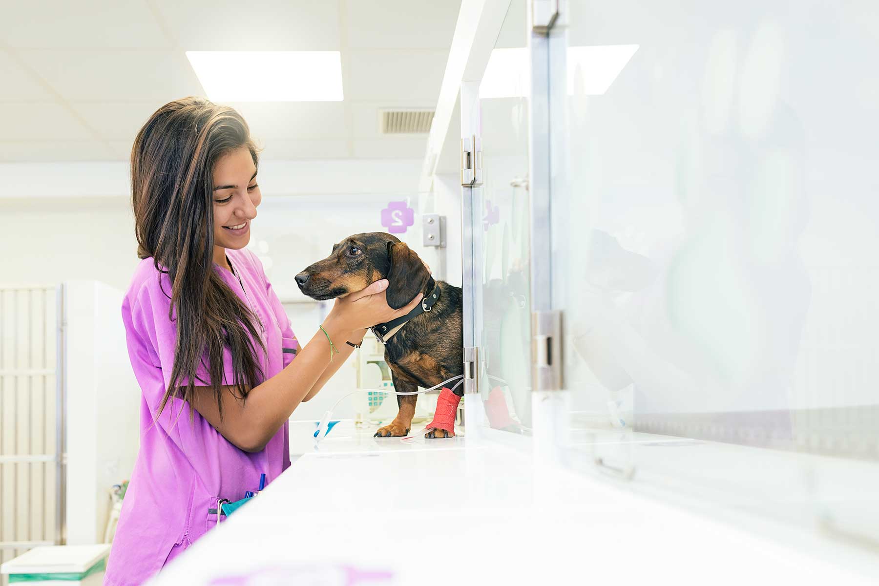
Finnbar first presented to us late one evening after having been lethargic, off his food and vomiting for 24 hours. During examination by one of our veterinarians, Finnbar was found to be uncomfortable in his abdomen and mildly dehydrated.
Xrays were performed that evening and revealed that Finnbar likely had a diaphragmatic hernia (a small hole in the muscular sheet that separates the chest and abdominal cavities) which was allowing a small portion of the liver to enter his chest. Diaphragmatic hernias can be congenital, however, can also form following traumatic injury (eg after being hit by a vehicle). In Finnbar’s case, there was no history of previous trauma that his owners were aware of. The X-rays also showed suggestive signs of a blockage in his intestine, but given this could not be confirmed from X-rays alone and that his hernia required further investigation to see whether it was causing a clinical problem, it was decided that the best next step would be to send him for a specialist abdominal ultrasound the following day.
The specialist ultrasonographer confirmed that Finnbar’s diaphragmatic hernia was most likely congenital and that although he did have a portion of liver poking through the hole into his chest, the liver appeared to be otherwise very healthy and was unlikely to be causing Finnbar any discomfort. In a way, the liver was also helping to “seal” the defect in his diaphragm, therefore preventing air from escaping from his chest cavity into his abdomen. The specialist ultrasonographer also confirmed that a roughly 5cm long piece of foreign material was present and causing a blockage in the small intestine. The most likely culprit based on its appearance was a trichobezoar (otherwise known as a hairball). Surgery was recommended to remove the blockage and allow Finnbar’s intestines to function normally again. Finnbar’s owner gave us the go-ahead to proceed with the surgery.
Surgery was performed successfully that day, with a large hairball being removed, and the rest of his gastrointestinal tract appearing otherwise normal at the time of surgery. After a 24-hour stay in hospital receiving further supportive care, Finnbar started eating normally again, was no longer vomiting and returned to his normal, extremely affectionate self. He was discharged home for further monitoring.
After initially recovering very well from surgery, five days later Finnbar became unwell again. Concerningly, he was showing very similar signs to those present when he first presented to the hospital. When we examined Finnbar, he was painful in his tummy again and some foreign material could be felt in his intestine.
Finnbar returned to the specialist ultrasonographer, who confirmed that a new furball (that was not present at the time of his last ultrasound) was now causing a blockage in a different part of his small intestine. Presumably, he had ingested enough fur while grooming himself over the previous five days to create a new furball large enough to cause a blockage. This made Finnbar one of the unluckiest cats we have ever met!
Given that Finnbar was becoming more unwell very rapidly, the difficult decision was made to take him to surgery again for the second time in a week. His second surgery also went very well. After the hairball was removed, Finnbar remained in hospital for another 24 hrs, where once again rallied very quickly. We have certainly had very few feline patients who flip over onto their backs for a cuddle as soon as you approach their cage!
In order to try and prevent another hairball ever developing, Finnbar also received a full body clip prior to being sent home and will continue to have regular haircuts long term. Although he was not particularly impressed by this, both the veterinarians and his owners agree that if it means Finnbar will never need another major surgery, then it will be worth the effort! We also think he still looks incredibly handsome in his more aerodynamic form!